BACTERIAL OCULAR INFECTIONS
Bacterial infections of the eye are common and distressing infectious disease in children and adults. They are a major cause of ocular morbidity in a large part of the population, and if left untreated, could lead to loss of vision.
The ocular infections can be classified as:-
• External Ocular Infections
• Intra Ocular Infections
External Ocular Infections are confined only to the external ocular structures. These include:-
• Infections of the Eyelids
• Infections of the Cornea
• Infections of the Lacrimal apparatus
EXTERNAL OCULAR INFECTIONS
A. INFECTIONS OF EYELIDS
• Conjunctivitis
Conjunctivitis is an acute inflammation of the conjunctiva (the outermost layer of the eye and the inner surface of the eyelids), most commonly due to an allergic reaction or an infection (usually viral, but sometimes bacterial).
It can be either by cause or by extent of the inflamed area.
By cause:-
i) Allergic conjunctivitis
ii) Bacterial conjunctivitis
iii) Viral conjunctivitis
iv) Chemical conjunctivitis
v) Neonatal conjunctivitis is often defined separately due to different organisms
By extent of involvement
i) Blepharoconjunctivitis is the dual combination of conjunctivitis with blepharitis (inflammation of the eyelids).
ii) Keratoconjunctivitis is the combination of conjunctivitis and keratitis (corneal inflammation).
iii) Episcleritis is an inflammatory condition that produces a similar appearance to conjunctivitis, but without discharge or tearing.
Signs and symptoms
Redness (hyperemia), irritation (chemosis) and watering (epiphora) of the eyes are symptoms common to all forms of conjunctivitis.
Allergic Conjunctivitis
Allergic conjunctivitis is typically itchy, sometimes distressingly so, and often involves some eye swelling. Chronic allergy often causes just itching or irritation.
Allergic conjunctivitis shows pale watery swelling or edema of the conjunctiva and sometimes the whole eyelid, often with a ropy, non-purulent mucoid discharge. There is variable redness.
Itching must be a primary symptom to make this diagnosis.
Bacterial Conjunctivitis
Bacterial conjunctivitis due to the common pyogenic (pus-producing) bacteria causes marked grittiness/irritation and a stringy, opaque, grey or yellowish mucopurulent discharge (mucus) that may cause the lids to stick together, especially after sleeping. Another symptom that could be caused by bacterial conjunctivitis is severe crusting of the infected eye and the surrounding skin. Bacteria such as Chlamydia trachomatis or Moraxella can cause a non-exudative but persistent conjunctivitis without much redness. The gritty and/or scratchy feeling is sometimes localized enough for patients to insist they must have a foreign body in the eye. The more acute pyogenic infections can be painful. Like viral conjunctivitis, it usually affects only one eye but may spread easily to the other eye. However, it is dormant in the eye for three days before the patient shows signs of symptoms.
Pyogenic bacterial conjunctivitis shows an opaque purulent discharge, a very red eye, and on bio microscopy there are numerous white cells and desquamated epithelial cells seen in the tear duct along the lid margin. The tarsal conjunctiva is a velvety red and not particularly follicular. Non-pyogenic infections can show just mild infection and be difficult to diagnose. Scarring of the tarsal conjunctiva is occasionally seen in chronic infections, especially in trachoma.
Viral Conjunctivitis
Viral conjunctivitis is often associated with an infection of the upper respiratory tract, a common cold, and/or a sore throat. Its symptoms include watery discharge and variable itch. The infection usually begins with one eye, but may spread easily to the other.
Viral conjunctivitis, commonly known as "pink eye", shows a fine diffuse pinkness of the conjunctiva which is easily mistaken for the "ciliary injection" of iritis, but there are usually corroborative signs on microscopy, particularly numerous lymphoid follicles on the tarsal conjunctiva, and sometimes a punctate keratitis.
Chemical Conjunctivitis
Irritant or toxic conjunctivitis is irritable or painful when the infected eye is pointed far down or far up. Discharge and itch are usually absent. This is the only group in which severe pain and discomfort may occur.
Irritant or toxic conjunctivitis show primarily marked redness. If due to splash injury, it is often present only in the lower conjunctival sac. With some chemicals, above all, with caustic alkalis such as sodium hydroxide—there may be necrosis of the conjunctiva with a deceptively white eye due to vascular closure, followed by sloughing of the dead epithelium. This is likely to be associated with slit-lamp evidence of anterior uveitis.
• Blepharitis
Blepharitis is an ocular condition characterized by chronic inflammation of the eyelid, the severity and time course of which can vary. It can onset acutely resolving without treatment within 2–4 weeks (this can be greatly reduced with lid hygiene) but more generally is a long standing inflammation varying in severity.
Signs and symptoms
Signs and symptoms that are associated with the chronic inflammation can be;
• Redness of the eyelids.
• Flaking of skin on the lids.
• Crusting at the lid margins, this is generally worst on waking.
• Cysts at the lid margin (hordeolum).
• Red eye.
• Debris in the tear film, seen under magnification (improved contrast with use of fluorescein drops).
• Gritty sensation of the eye.
• Reduced vision.
Common signs and symptoms of blepharitis also include itching, irritation and burning as well as a foreign body sensation. Some patients experience eye dryness, which can cause a certain degree of discomfort.
Eye redness and swelling tend to appear in more advanced cases, and they are rarely primary symptoms. The symptoms can slightly vary based on the exact cause of the condition. Blepharitis due to an allergy can cause dark lids, symptom which is known as "allergic shiner" and which tends to be more frequent in children rather than adults. Infectious blepharitis is accompanied by a yellow or green-colored discharge which is more abundant in the morning and which leads to stuck lids. Blepharitis may also cause eyelid matting or "gluing" of the lashes. Other blepharitis symptoms include sensitivity to light, eyelashes that grow abnormally or even loss of eyelashes. Also, the tears might seem frothy or bubbly in nature and mild scarring might occur to the eyelids. The symptoms and signs of blepharitis are often erroneously ascribed by the patient as being due to "recurrent conjunctivitis".
Staphylococcal Blepharitis
Staphylococcal blepharitis is caused by infection of the anterior portion of the eyelid by Staphylococcal bacteria. As the infection progresses, the sufferer may begin to notice a foreign body sensation, matting of the lashes, and burning. Usually, the primary care physician will prescribe topical antibiotics for staphylococcal blepharitis, as this is an acute condition and should heal quickly. The condition can sometimes lead to a chalazion or a stye.
• Hordeolum / Stye
An external stye or hordeolum is an infection of the sebaceous glands of Zeis at the base of the eyelashes, or an infection of the apocrine sweat glands of Moll. External styes form on the outside of the lids and can be seen as small red bumps. Internal styes are infections of the meibomian sebaceous glands lining the inside of the eyelids. They also cause a red bump underneath the lid with only generalized redness and swelling visible on the outside. Styes are similar to chalazia, but tend to be of smaller size and are more painful and usually produce no lasting damage. Styes are characterized by an acute onset and usually short in duration (7-10 days without treatment) compared to chalazia that are chronic and usually do not resolve without intervention.
Causes
Styes are commonly caused by a Staphylococcus aureus bacterial infection, or by the blocking of an oil gland at the base of the eyelash. Although they are particularly common in infants, styes are experienced by people of all ages. Styes can be triggered by poor nutrition, sleep deprivation, lack of hygiene or rubbing of the eyes. Sharing of washcloths or face towels should be curtailed to avoid spreading the infection between individuals. Styes can last from 1 to 2 weeks without treatment, or as little as 4 days if treated properly.
Signs and Symptoms
The first sign of a stye is a small, yellowish spot at the center of the bump that develops as pus expands in the area.
Other stye symptoms may include:
• A lump on the top or bottom eyelid
• Localized swelling of the eyelid
• Pain which becomes more intense if the pus ruptures
• Redness
• Tenderness to touch
• Crusting of the eyelid margins
• Burning in the eye
• Droopiness of the eyelid
• Scratchy sensation on the eyeball
• Blurred vision
• Mucous discharge in the eye
• Irritation of the eye
• Light sensitivity
• Tearing
• Discomfort during blinking
• Sensation of a foreign body in the eye.
• Chalazion
A chalazion also known as a meibomian gland lipogranuloma is a cyst in the eyelid that is caused by inflammation of a blocked meibomian gland, usually on the upper eyelid. Chalazia differ from styes (hordeola) in that they are subacute, nontender, and usually painless nodules. May become acutely inflamed but, unlike a stye, usually point inside the lid rather than on the lid margin. A chalazion or meibomian cyst could take months to fully heal with treatment and could take years to heal without any major complications.
Signs and symptoms
• Swelling on the eyelid
• Eyelid tenderness
• Sensitivity to light
• Increased tearing
• Heaviness of the eyelid
• Meibomianitis
Inflammation of the little glands called Meibomian glands located in the eyelids that make a lubricant which they discharge through their tiny openings in the edges of the lids. The lubricant is a fatty substance called sebum characteristic of sebaceous glands. Meibomianitis may be due to allergy, acne in adolescence, or rosacea. Chronic inflammation of the Meibomian glands leads to cysts, called chalazions. Treatment usually consists of lubricant eye drops, warm compresses to the eyes, and careful cleansing of the eyelids. An antibiotic ointment may be prescribed to apply to the edge of the eyelid.
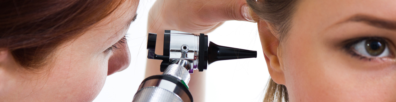
B. INFECTION OF CORNEA
• Keratitis
Keratitis is the medical term for inflammation of the cornea. The cornea is the dome-shaped window in the front of the eye. When looking at a persons eye, one can see the iris and pupil through the normally clear cornea. The cornea bends light rays as a result of its curved shape and accounts for approximately two-thirds of the eyes total optical power, with the lens of the eye contributing the remaining one-third. Only the very thin tear film lies between the front of the cornea and our environment.
The cornea is about 0.5 millimeter thick. The back of the cornea is bathed in the aqueous fluid that fills the anterior chamber of the eye. The cornea has a diameter of about 13 millimeters (½ inch) and, together with the sclera (the white part of the eye) forms the entire outer coat of the eye.
Causes of Keratitis
Keratitis, the eye condition in which the cornea becomes inflamed, has many potential causes. Various types of infections, dry eyes, injury, and a large variety of underlying medical diseases may all lead to keratitis. Some cases of keratitis result from unknown factors.
Different types of Keratitis
Keratitis can be classified by its location, severity, and cause.
• If keratitis only involves the surface (epithelial) layer of the cornea, it is called superficial keratitis. If it affects the deeper layers of the cornea (the corneal stroma), it is called stromal keratitis or interstitial keratitis . It may involve the center of the cornea or the peripheral part of the cornea (that portion closest to the sclera) or both. Keratitis may affect one eye or both eyes.
• Keratitis may be mild, moderate, or severe and may be associated with inflammation of other parts of the eye. Keratoconjunctivitis is inflammation of the cornea and the conjunctiva. Kerato-uveitis is inflammation of the cornea and the uveal tract, which consists of the iris, ciliary body, and choroid. Keratitis may be acute or chronic. It may occur only once or twice in an eye or be recurrent. It may be limited in its effects on the eye or be progressive in its damage.
• Infection is the most frequent cause of keratitis. Bacteria, viruses, fungi, and parasitic organisms may all infect the cornea, causing infectious or microbial keratitis.
A. Bacteria most frequently responsible for keratitis include Staphylococci, Hemophilus, Streptococci, and Pseudomonas. If the front surface of the cornea has been damaged by a small scratch and the surface is not intact, almost any bacteria, including atypical mycobacterium, can invade the cornea and result in keratitis. Ulcerations of the cornea may occur, a condition known as ulcerative keratitis. Before the advent of antibiotics, syphilis was a frequent cause of keratitis.
B. Viruses that infect the cornea include respiratory viruses, including the adenoviruses and others responsible for the common cold. The herpes simplex virus is another common cause of keratitis. The herpes zoster virus (the virus responsible for chickenpox and shingles) may also cause keratitis if shingles involve the forehead.
C. Fungi such as Candida, Aspergillus, and Nocardia are unusual causes of microbial keratitis, more frequently occurring in people who are immunocompromised because of underlying illnesses or medications. Fusarium keratitis, a type of fungal infection, occurs primarily in contact-lens wearers. Bacterial co-infection can complicate fungal keratitis.
D. Contact-lens wearers are also susceptible to acanthamoeba keratitis caused by an amebic parasite. "River blindness," or onchocercal keratitis, is another parasitic infection of the cornea, rarely seen in developed countries.
E. Physical or chemical trauma is a frequent cause of keratitis. The injury may become secondarily infected or remain noninfectious. Retained corneal foreign bodies are frequent sources of keratitis. Ultraviolet light from sunlight (snow blindness), a tanning light or a welders arc, contact-lens over wear, and chemical agents, either in liquid form splashed into the eye or in gases in the form of fumes can all result in noninfectious keratitis. Chemical injury or contact lens-related keratitis often causes superficial punctate keratitis, in which the examiner notices myriads of injured surface cells on the affected cornea.
F. Disturbances in the tear film may lead to changes in the corneal surface through drying of the corneal epithelium. This type of keratitis is usually superficial and most commonly is related to dry eyes and is known as keratitis sicca. If the eyes are extremely dry, the surface cells may die and form attached filaments on the corneal surface, a condition known as filamentary keratitis. Inability to close the eyelids properly can also lead to cornea drying, which a condition is termed exposure keratitis.
G. Allergies to airborne pollens or bacterial toxins in the tears may also cause a noninfectious type of keratitis. Autoimmune diseases create a similar appearance, often affecting the periphery of the cornea, termed marginal keratitis or limbic keratitis.